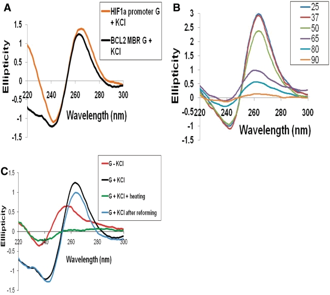
Figure 2.
CD studies on the MBR show formation of parallel, G-quadruplex structure on the template strand. (A) The BCL2 MBR and HIF-1α promoter G-strand (positive control) were incubated in presence of 100 mM KCl and TE at 37°C for 1 h and the CD spectra was taken using JASCO J-810 spectropolarimeter with a scan range of 220–300 nm. (B) CD spectra for the MBR template strand in TE with KCl at different temperatures. The various temperatures used are 25, 37, 50, 65, 80 and 90°C. (C) CD spectra for the MBR template strand in TE with (G + KCl) or without KCl (G − KCl), or same after heating to 90°C (G + KCl + heating) and then incubation at 37°C overnight (G + KCl after reforming), in order to reform the structure.

