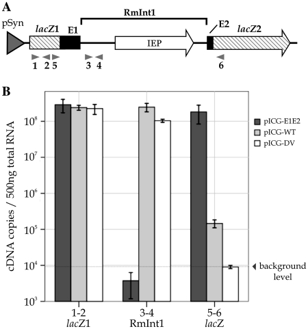
Figure 4.
Quantitative analysis of RmInt1 in S. meliloti. (A) Map of the lacZ region of pICG-WT. The RmInt1 intron with the IEP (white arrow) encoded in DIV and minimal flanking sequences from the ISRm2011-2 insertion sequence (E1 and E2) (black boxes) are located in the lacZ gene (hatched arrow). The interrupted lacZ gene is shown as lacZ1 and lacZ2 exons. Arrows under the map indicate the primers used in the real-time RT–PCR assay. Primer pair 1–2 was used to amplify the lacZ1; primers 3–4 were used to amplify the RmInt1 intron and primers 5–6 were used to amplify the ligated β-galactosidase gene lacZ. (B) Total RNA was isolated from S. meliloti containing the wild-type intron pICG-WT, the intron-less construct pICG-E1E2 and the splicing-defective mutant pICG-DV. The levels of lacZ1 (1–2), RmInt1 intron (3–4) and ligated β-galactosidase gene lacZ (5–6) mRNA were determined by an absolute quantification method. Each measurement is the mean of at least three independent RNA preparations.

