Fig. 1.
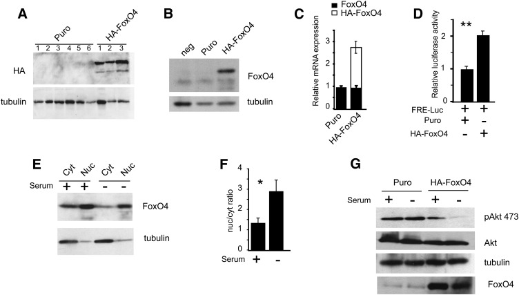
Derivation of stable 3T3L1 fibroblasts expressing a hemaglutinin-tagged (HA) form of FoxO4. A: Western blot analysis of protein extracts from stable cell lines transduced with pBabe Puro or pBabe HA-FoxO4 retroviruses. HA-FoxO4 was detected with anti-HA, while anti-tubulin was used to reveal protein loading. The Western blot shows six negative and three positive cell lines. B: Western blot with anti-FoxO4 from protein extracts of two clones, a Puro negative control, and HA-FoxO4. The protein band below HA-FoxO4 is likely to represent endogenous FoxO4 and appears in all three lanes. C: Expression levels by real-time qPCR of endogenous FoxO4 and HA-FoxO4 mRNA in Puro and HA-FoxO4 cell lines (n = 6 in each group). D: Cotransfection of FRE-luciferase with either Puro or HA-FoxO4. Relative luciferase activity is shown for three independent experiments. Asterisks indicate P < 0.01. E: Western blot of nuclear and cytoplasmic protein extracts from HA-FoxO4 cells either serum fed (+) or starved (−). The blots were conjugated with either anti-FoxO4 or anti-tubulin. A small amount of cytoplasmic tubulin is carried over to the nuclear fraction. F: Nuclear-to-cytoplasmic (nuc/cyt) ratio of HA-FoxO4 in serum fed (+) and starved (−) conditions as determined by densitometric scan of the Western blot shown in E. Asterisk indicates P < 0.05. G: Western blot analysis of protein lysates from Puro and HA-FoxO4 cells either serum fed (+) or starved (−) and conjugated to anti-phosphoserine 473 Akt, anti-Akt, anti-tubulin (for protein loading control), or anti-FoxO4.

