Fig. 4.
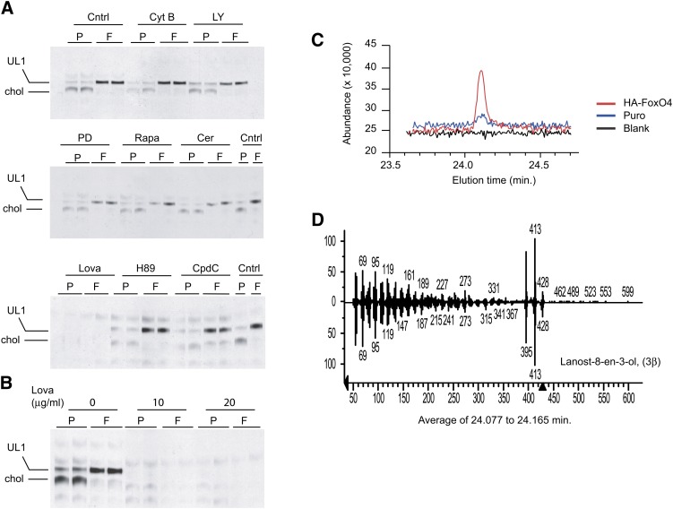
Characterization of the HA-FoxO4 UL1 lipid. A: TLC plate autoradiographs of lipid extracts from Puro (P) and HA-FoxO4 (F) fibroblasts either untreated (Cntrl) or pretreated with 10 μM cytochalasin B (Cyt B), 50 μM LY294002 (LY), 50 μM PD 98059 (PD), 0.1 μM rapamycin (Rapa), 10 μM cerulenin (Cer), 20 μg/ml lovastatin (Lova), 5 μM H89, or 20 μM compound C (CpdC) prior to 14C-acetate labeling. Lipids are labeled as cholesterol (chol) and the FoxO4 unknown lipid as UL1. Each lane represents a lipid extract from two combined wells of a 24-well plate. Inhibition of cholesterol and UL1 are seen with lovastatin treatment. B: TLC plate autoradiograph of lipid extracts from 14C-labeled Puro and HA-FoxO4 cells treated with two different concentrations of the HMG-CoA reductase inhibitor lovastatin (Lova). C: GC and elution times of blank, Puro, and HA-FoxO4 lipid extracts. D: MS fingerprint of UL1, which consists of lanost-8-en-3-ol (3β), commonly called DHL.

