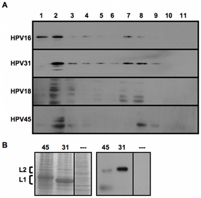
Figure 4. Western blot analysis of Opriprep-purified L1+L2 VLPs.
(A) HPV16, HPV31, HPV18, and HPV45 VLPs were Optiprep-fractionated and fractions were assayed for L2 content by probing Western blots with anti-HPV16 L2 polyclonal antibody anti-P56/75 #1. (B) Side-by-side SDS-PAGE gels run with equivalent amounts of HPV45 and HPV31 L1+L2 VLPs from Optiprep fraction #8 were either Coomassie-stained, or Western blotted with anti-P56/75 #1. L2 and L1 bands are indicated by brackets (note the small size difference in L2 and the larger size difference in L1). A control lane that represents an SDS-PAGE gel of Optiprep-purified 293TT cell lysate without capsid protein was also included for the Coomassie stained samples (—).

