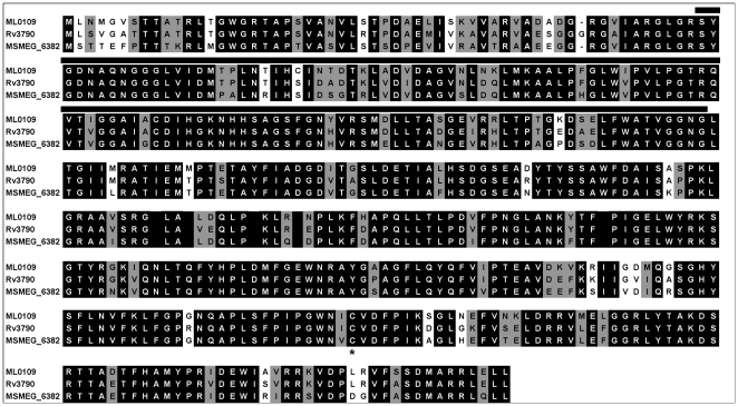
Figure 2. DprE1 alignment.
Homologs from M. leprae (ML0109), M. tuberculosis (Rv3790) and M. smegmatis (MSMEG_6382) were aligned using CLUSTALW [25]. Residues that are completely conserved are reverse shaded while similar residues are indicated in grey. The putative FAD-binding domain is shown by a solid line. The conserved cysteine residue that is altered in BTZ-resistant mycobacteria [11] is indicated by an asterix.

