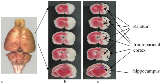
Figure 2. Brain infarction revealed by triphenyltetrazolium chloride (TTC) staining.
(a) Rat brain (b) original TTC image of brain infarction after 90 minutes middle cerebral artery occlusion; and (c) image with labels. TTC reacts with dehydrogenases in viable cells and results in a “brick-red” color, and the white area indicates the infarction. It was clearly detected that ipsilateral motor cortex and striatum were affected with middle cerebral artery occlusion.

