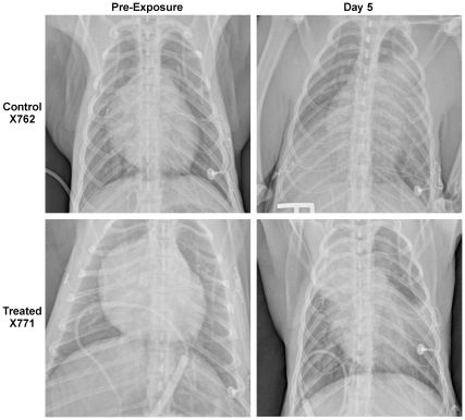
Figure 4. Chest radiographs of a control and treated animal prior to and 5 days after challenge.
Chest radiographs obtained prior to challenge (left) and 5 days post exposure (right) for two macaques typical of findings in other animals, The untreated control animal (X762) displayed large infiltrates in the right caudal, left middle and left cranial lobes immediately prior to euthanasia for respiratory distress. The treated animal (X771) had initiated levofloxacin therapy 20 hours prior to pulmonary imaging on day 5 pe and displayed large infiltrates in the right cranial lobe and left caudal lobe, superior segment, with minor infiltrates in the left cranial lobe and right caudal lobe.

