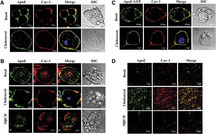
Fig. 1.
Cellular localization of adipocyte apoE and cav-1 under basal and CH-enriched conditions. Adipocytes were incubated in 100 μM BSA/DMEM or this medium containing 10 μg/ml CH for 2 h or 10 mM MβCD for 50 min as indicated. A: Cultures of primary rat adipocytes were stained with anti-apoE or anti-cav-1 and used for confocal microscopy as described in Methods. B: 3T3-L1 adipocytes were stained with anti-apoE or anti-cav-1 and used for confocal microscopy as described in Methods. C: Endogenously synthesized apoE was detected in 3T3-L1 cells after expression of an apoE-GFP protein. Cells were also stained with anti-cav-1 as described in Methods. D: Plasma membrane sheets were isolated (as described in Methods) after incubation of intact 3T3-L1 cells under basal conditions, with CH, or with MβCD. Plasma membrane sheets were stained with anti-apoE and anti-cav-1 and imaged with confocal microscopy.

