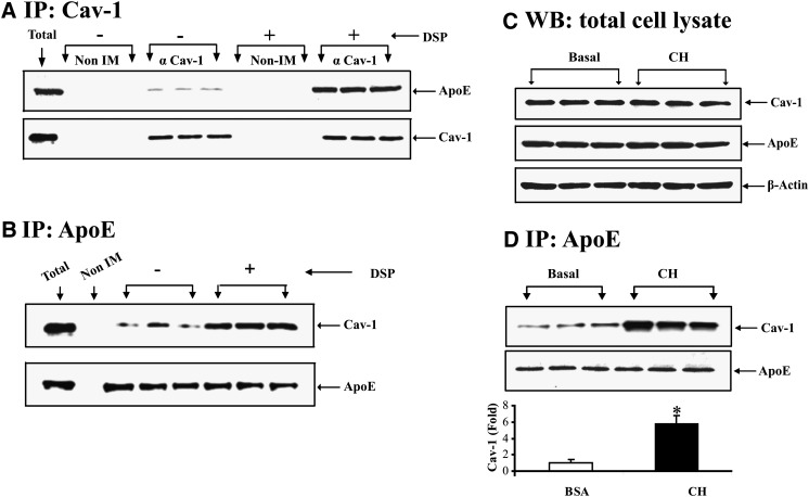
Fig. 2.
Coimmunoprecipitation of apoE and cav-1. A: 3T3-L1 cells were incubated in 100 μM BSA for 2 h. After this incubation, cells were lysed in nondenaturing buffer and the lysate was for immunoprecipitation using nonimmune serum or anti-cav1 antibody as indicated. Where indicated, 3T3-L1 cells were treated with the cell permeant reducible cross-linker DSP prior to lysis in denaturing but nonreducing buffer. The immunoprecipitates were eluted from protein G agarose beads in denaturing buffer containing 5% BME to cleave the cross-linker. The eluates were then separated on an SDS-PAGE and utilized for Western blot of cav-1 and apoE. B: 3T3-L1 cells were treated with the cell permeant reducible cross-linker DSP as described in Methods. Cells were lysed using denaturing conditions (but without reducing agents) and immunoprecipitated with an antibody to apoE. The immunoprecipitates were eluted from protein G agarose beads in denaturing buffer containing 5% BME to cleave the cross-linker. The eluates were then separated on SDS-PAGE and utilized for Western blot of cav-1 and apoE. C: 3T3-L1 cells were incubated in 100 μM BSA alone, or this medium plus 10 μg/ml CH for 2 h. After this incubation, cell lysates were used for Western blot for apoE and cav-1 as indicated. D: 3T3-L1 cells were incubated in 100 μM BSA alone, or this medium plus 10 μg/ml CH for 2 h. After this incubation, cells were lysed in nondenaturing buffer and immunoprecipitated with an antibody to apoE. The immunoprecipitates were eluted from protein G agarose beads using denaturing and reducing conditions. The eluates were separated on SDS gels and used for Western blot for cav-1 and apoE.

