
Figure 1.
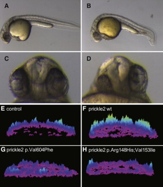
Zebrafish Prickle Mutations Orthologous to Those Identified in Human Epilepsy Patients Show Altered Activity in CE and Calcium Release
(A–D) Morphological phenotypes at 28 hpf. Compared to wild-type uninjected embryos (A), prickle2-injected embryos (B) display a shorter A-P axis and a kinky tail. The lateral view is shown, and anterior is to the left. Moreover, prickle2-injected embryos (D) show defects in eye and forebrain patterning at 28 hpf. (C) Uninjected embryo. Ventral view. The numbers of embryos with defects in each RNA-injected group are shown in Table 2.
(E–H) Surface plots of calcium release activity were generated from images of live zebrafish embryos. The height and color of the peaks indicates the number of calcium fluxes observed over the course of the experiment; the embryos are oriented in a lateral position. (E) A control injected embryo. (F) An embryo overexpressing wild-type prickle2 RNA. (G) An embryo overexpressing prickle2Val605Phe-mutant-encoding RNA. (H) An embryo overexpressing prickle2 Arg148His; Val153Ile-mutant-encoding RNA.