
Figure 5.
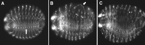
Neuronal Defects Are Observed in prickle Mutant Embryos
Images (20×) of control 14–16 hr yw67 embryos (A) are compared to two 14–16 hr pksple1homozygous mutant embryos (B and C) that have been stained with the 22C10 antibody so that PNS neurons are visualized. Note the location of chordotonal organs (asterisks in A) as well as the location of the ventral nerve chord (upward arrow in A) in the control embryo. For the mutant embryo in (B), note the aberrant neuronal processes that have joined (arrow) as well as the abnormal location of two chordotonal organs (asterisks). For the mutant embryo in (C), note the overall disorganized PNS staining pattern, as well as the improperly positioned neurons associated with the chordotonal organ in abdominal segment 6 (asterisk). Anterior is to the left, and ventral is down but slightly rotated toward the viewer.