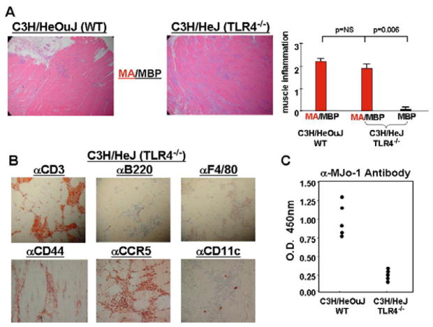
Figure 5. Induction of myositis is not dependent on TLR4 signaling.
In Panel A, photomicrographs (100×) correspond to H&E-stained muscle tissue obtained from C3H/HeOuJ (WT) versus C3H/HeJ (TLR4−/−) mice 17 days following IM immunization with MA/MBP. The bar graph indicates the relative severity of muscle inflammation resulting from IM immunization with MA/MBP (C3H/HeOuJ (n=10), C3H/HeJ (n=20)) or MBP (C3H/HeJ (n=5)). Immunohistochemical staining (400×) with antibodies targeting CD3, B220, F4/80, CD44, CCR5, and CD11c reveals the relative number as well as activation state of T cells, B cells, macrophages, and dendritic cells comprising inflammatory infiltrates of muscle tissue obtained from MA/MBP-immunized C3H/HeJ mice (Panel B). Finally, the ELISA data in panel C demonstrate relative anti-MJo-1 antibody formation in C3H/HeOuJ versus C3H/HeJ mice immunized with MA/MBP. Data points correspond to mean OD450 values generated by 1:1000 dilutions of sera from individual mice.

