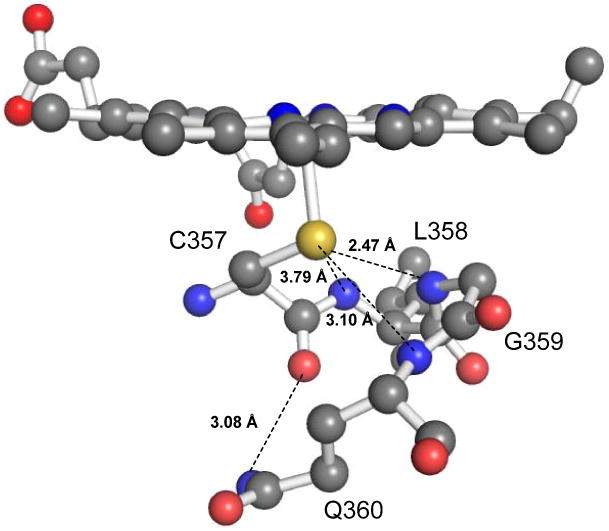
Figure 1.

Proximal hydrogen bonding network of Cytochrome P450cam. The orange, yellow and blue balls represent the heme iron, the sulfur atom and the main chain amide nitrogen atoms of Leu358, Gly359, and Gln360, respectively. The dashed lines represent the NH⋯S hydrogen bonds in the Cys pocket. The image was generated using PyMOL from PDB code 2CPP.(35)
