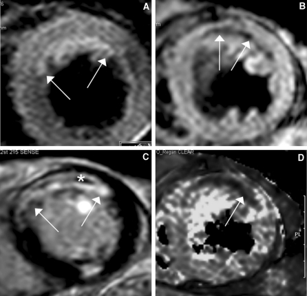
Fig. 1.
Example of a 76 year old male patient with an acute myocardial infarction (STEMI) of the anterior wall after acute PCI of the occluded LAD with stent implantation (pain-to-balloon time 150 min.). a The water sensitive T2-STIR image demonstrates the edema in the anterior wall (white arrows), c the LGE images demonstrate an almost transmural myocardial infarction (white arrows) with central MVO (asterisk). This indicates almost no myocardial salvage after successful revascularization of the LAD. b shows the T2* image at TE = 15 ms and d the T2* imaging map of the T2* mapping. The central dark area (white arrow) represents pixels with a T2* decay <20 ms indicating postreperfusionhemorrhage

