Fig. 4.

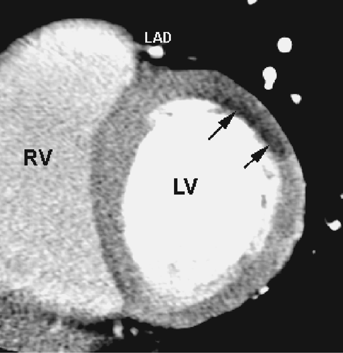
Example of acute MI due to occlusion of an obtuse marginal artery [126]. First pass MSCT shows local hypoenhancement of the antero-lateral wall. This perfusion defect was leading to find the culprit lesion in a 3 vessel disease patient

Example of acute MI due to occlusion of an obtuse marginal artery [126]. First pass MSCT shows local hypoenhancement of the antero-lateral wall. This perfusion defect was leading to find the culprit lesion in a 3 vessel disease patient