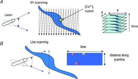
Figure 5. Application of confocal microscopy to Ca2+ imaging in living cells.

Cells are loaded with a fluorescent Ca2+ indicator (e.g. fluo-4) and excited with a scanning laser. A, in XY scanning mode a single image plane is scanned repeatedly, allowing any [Ca2+]i rises to be recorded and their location identified. B, line scanning greatly increases the speed of acquisition by imaging only along a single line rather than a full XY plane. The resulting images show how [Ca2+]i changes locally at each point on the line as time passes. This is particularly useful when imaging fast events, such as Ca2+ sparks.
