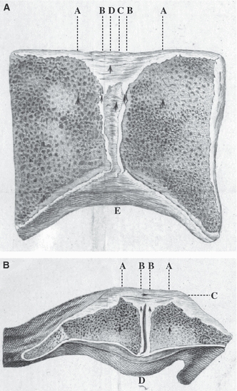
Fig. 1.

Sections of the pubic symphysis as depicted by William Hunter (1762). (A) Coronal section from a nulliparous female. AA, pubic bone; BB, cartilage; C, interior ‘ligamentous substance’; D, superior pubic ligament; E, inferior pubic ligament. (B) Axial section from a woman with puerperal fever. AA, pubic bone; BB, cartilage; C, interior ‘ligamentous substance’, the anterior pubic ligament that blends with tendinous fibres from adjacent muscle attachments; D, posterior pubic ligament that projects prominently in some subjects. The interpubic cleft is visible in the centre of the joint.
