
Fig. 3.
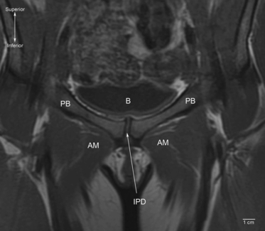
Coronal T1-weighted magnetic resonance image of the pubic symphysis in a supine 33-year-old woman (anterior view). Note that the joint is asymmetric. B, bladder; AM, adductor muscle group; PB, pubic bone; IPD, interpubic disc.
Official websites use .gov
A
.gov website belongs to an official
government organization in the United States.
Secure .gov websites use HTTPS
A lock (
) or https:// means you've safely
connected to the .gov website. Share sensitive
information only on official, secure websites.
Coronal T1-weighted magnetic resonance image of the pubic symphysis in a supine 33-year-old woman (anterior view). Note that the joint is asymmetric. B, bladder; AM, adductor muscle group; PB, pubic bone; IPD, interpubic disc.