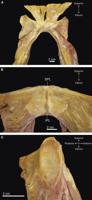
Fig. 4.

Dissection images of the pubic symphysis from an elderly cadaver of unknown parity. (A) Anterior view of the pubic symphysis showing blending of the tendons of rectus abdominis (RA) and pyramidalis (P) with the anterior pubic ligament. Note the decussation of the gracilis (G) tendons. AL, adductor longus. (B) Posterior view of the superior (SPL) and inferior (IPL) pubic ligaments. (C) The left medial pubic surface after bisection of the fibrocartilaginous interpubic disc.
