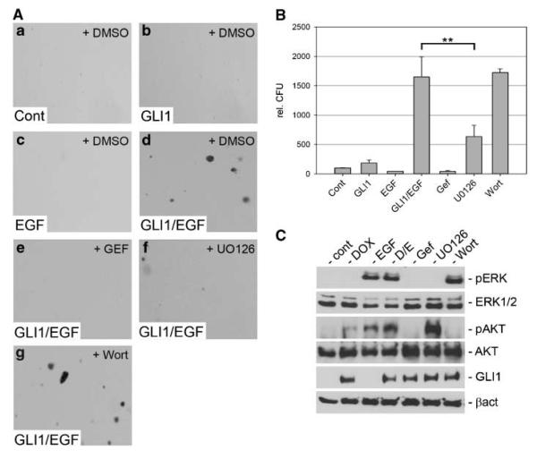
Figure 3.
Anchorage-independent growth of HaCaT keratinocytes by combined activation of GLI1 and EGFR signaling. A, soft agar cultures of control HaCaT keratinocytes (no EGFR, no GLI1; a), keratinocytes expressing GLI1 only (b), EGF (10 ng/mL) treated keratinocytes (c), or HaCaT keratinocytes expressing GLI1 and treated with EGF (d). e-g, HaCaT cells with activated EGFR and GLI1 treated with gefitinib (Gef; e), UO126 (f), or wortmannin (g). B, quantification of assays shown in A. Statistical analysis was done by Student's t test. **, P < 0.005. Data represent the mean value of three independent experiments, each performed in triplicate. C, Western blot analysis of doxycycline-inducible GLI1 HaCaT keratinocytes showing specific activation and inhibition of MEK/ERK and PI3K/AKT function by treatment with the respective compounds. Samples Gef, UO126, and Wort were also treated with doxycycline and EGF. Cont, control; DOX, doxycycline; D/E, doxycycline/EGF treated; Wort, wortmannin.

