Figure 4.
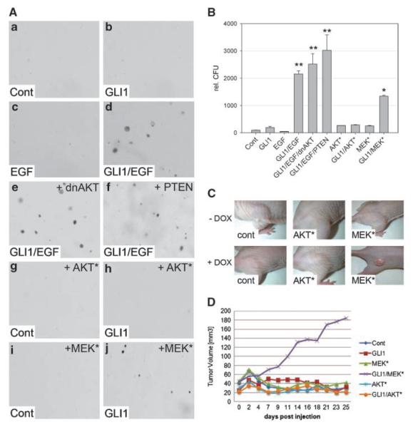
MEK but not AKT synergizes with GLI1 in oncogenic transformation. A, HaCaT keratinocytes grown in soft agar either in the absence of both EGF and GLI1 expression (a), in the presence of GLI1 (b), in the presence of EGF (c), or in the presence of both GLI1 and EGF (d). Anchorage-independent growth is not affected by coexpression of dnAKT (e) or PTEN (f). HaCaT cells expressing constitutively active AKT (AKT*) in the absence (g) or presence of GLI1 (h). HaCaT cells expressing dominant-active MEK (MEK*) alone (i) or in combination with GLI1 (j). B, quantitative analysis of soft agar cultures shown in A. Statistical analysis was done by Student's t test. **, P < 0.001; *, P < 0.005. Data represent the mean value of three independent experiments, each performed in triplicate. C, nude mice (n = 8 for each cell line) injected with doxycycline-inducible GLI1 HaCaT keratinocytes expressing either AKT* or MEK*. GLI1 expression was induced by doxycycline in drinking water (+ DOX). D, quantitative analysis of tumor growth in nude mice shown in C.

