Figure 1.
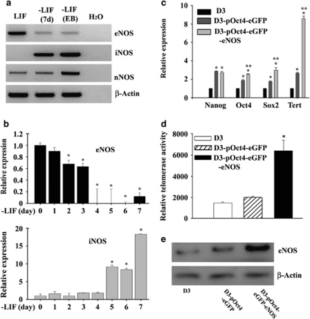
Differential expression of nitric oxide (NO) synthases and expression of self-renewal markers in mESCs. (a) NO synthase expression during differentiation. Sorted D3-pOct4-eGFP mESCs were cultured for 7 days in the presence or absence of LIF attached to a surface and for 14 days by hanging drop protocol (EB preparation). eNOS, iNOS and nNOS expression were detected by RT-PCR. The images are representative of three experiments. (b) Time course of eNOS and iNOS expression. R1-E mESCs were cultured in the absence of LIF for 7 days. eNOS and iNOS expression were detected by qRT-PCR, β-actin was used as an endogenous control and cycle threshold (Ct) values were normalized with respect to those of cells cultured in the presence of LIF (day 0). Data shown are from three independent experiments. *P≤0.005 with respect to day 0. (c) qRT-PCR of undifferentiation markers. D3 mESCs, D3-pOct4-eGFP mESCs and D3-pOct4-eGFP-eNOS mESCs were cultured in the presence of LIF and gene expression was determined by qRT-PCR. Data are the mean ± S.E.M. of three experiments; *P≤0.005 when compared with D3 cells and **P≤0.005 when compared with D3-pOct4-eGFP cells. (d) Telomerase activity in D3 mESCs, D3-pOct4-eGFP mESCs and D3-pOct4-eGFP-eNOS mESCs. Each data point represent the mean ± S.E.M. of three experiments; *P≤0.005 when compared with other cell types. (e) Western blot analysis of eNOS in D3 mESCs, D3-pOct4-eGFP mESCs and D3-pOct4-eGFP-eNOS mESCs. Representative image of three independent experiments

