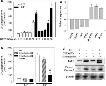
Figure 2.
Effect of different NO concentrations on apoptosis and cell survival. (a) Effect of DETA-NO on DNA fragmentation. D3-pOct4-eGFP mESCs were cultured for 7 days in the presence or absence of LIF treated with different concentrations (2–50 μM) of DETA-NO. DNA fragmentation was measured. Data are the mean ± S.E.M. of five independent experiments. *P≤0.005 versus. D3 mESCs cultured in the presence of LIF and in the absence of DETA-NO (b) D3, D3-pOct4-eGFP and D3-pOct4-eGFP-eNOS mESCs were cultured in the presence or absence of LIF for 7 days and DNA fragmentation was measured. Data are the mean ± S.E.M. of five independent experiments. *P≤0.005 versus. D3 and D3-pOct4-eGFP mESCs cultured in the absence of LIF. **P≤0.005 versus. mESCs cultured in the presence of LIF. (c) Analysis of pro-apoptotic and anti-apoptotic gene expression in D3-pOct4-eGFP mESCs. Cells were cultured in the absence of LIF and treated with 2 μM DETA-NO gene expression was determined by qRT-PCR. β-actin was used as an endogenous control and Ct values were normalized with respect to the Ct of cells cultured in absence of LIF. Data are the mean ± S.E.M. of three experiments. (d) Effect of DETA-NO on PARP and caspase 3 cleavage. D3-pOct4-eGFP mESCs were cultured in the presence or absence of LIF for 7 days and supplemented with 2 μM DETA-NO. PARP and cleaved caspase 3 were detected by western blot. The positive control for apoptosis was 1 μM Staurosporine. Figures are representative of three independent experiments

