Figure 4.
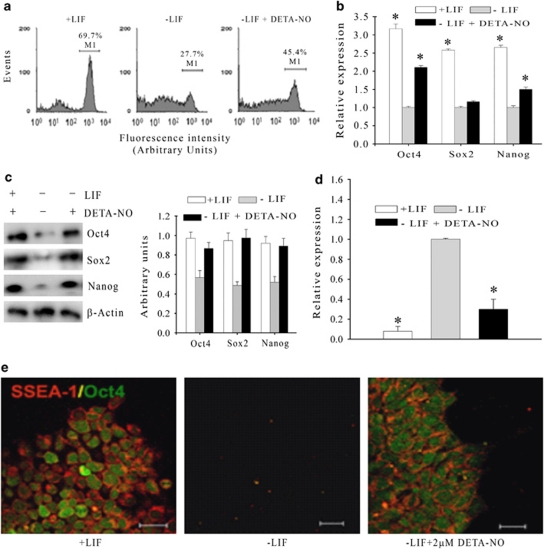
Exogenous NO arrests the differentiation induced by LIF withdrawal in mESCs. (a) D3-pOct4-eGFP mESCs from passage 10 after sorting were cultured for 7 days in the presence (left panel) or absence (medium panel) of LIF and in the absence of LIF plus 2 μM DETA-NO (right panel). GFP intensity was measured by flow cytometry. Cells with fluorescence intensity similar or higher than that observed for mESCs transfected with the pOct4-eGFP construct and cultured for 7 days in the presence of LIF were considered positive for eGFP (M1 population). Each graph is representative of five independent experiments (n=5). (b) qRT-PCR analysis of Oct4, Sox-2 and Nanog in D3-pOct4-eGFP mESCs cultured for 7 days under the indicated conditions. β-actin was used as the endogenous control and Ct values were normalized with respect to the Ct of cells cultured in the absence of LIF. Data are the mean ± S.E.M. of three experiments. *P≤0.005 versus. cells cultured in the absence of LIF. (c) Western blot analysis of Oct4, Sox-2 and Nanog in D3-pOct4-eGFP mESCs cultured for 7 days under the indicated conditions. Representative images and densitometry quantification are from five independent experiments. (d) qRT-PCR analysis of Brachyury expression in D3-pOct4-eGFP mESCs. β-actin was used as the endogenous control and Ct values were normalized with respect to the Ct of cells cultured in the absence of LIF. Data are the mean ± S.E.M. of three experiments. *P≤0.005 versus. cells cultured in the absence of LIF. (e) Confocal images of cells positive for SSEA-1 in non-transfected D3 mESCs cultured for 7 days in the presence (left panel) or absence (middle panel) of LIF or in the absence of LIF plus 2 μM DETA-NO (right panel). Nuclei are counter-stained with DAPI. Images are from three independent experiments. Scale bars are 20 μm

