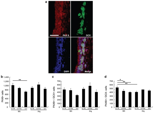
Figure 4.
Stem/progenitor cells in the SVZ of Hq mice after IR. Representative microphotographs of the SVZ (a) stained for PAX6 (red) and doublecortin (DCX) (green) 7 days after IR. Bar=25 μm. Quantification of the number of (b) the PAX6-positive cells, (c) the PAX6- and DCX-positive cells (PAX6+/DCX+) and (d) the PAX6-positive, DCX-negative cells (PAX6+/DCX−). Data represent mean±S.E.M. *P<0.05, **P<0.01, n=7 per group

