Figure 6.
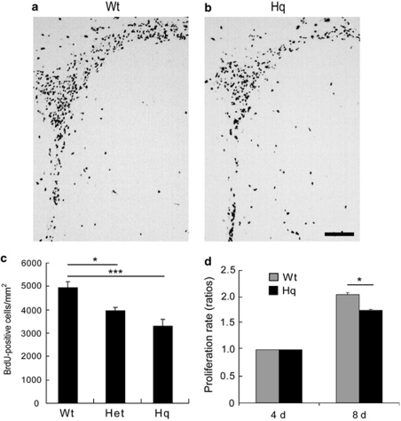
Cell proliferation in the SVZ of non-IR Hq mice. Representative microphotographs from Wt (a) and Hq (b) brains of the SVZ stained for BrdU 24 h after injection on P10, without IR. Bar=100 μm. (c) Quantification of the BrdU-positive cells in the SVZ of Wt (n=6), Het (n=6) and Hq (n=7). (d) Proliferation of cultured NSPCs from the SVZ of Wt and Hq mice 4 and 8 days after plating (n=10 per group). Data represent mean±S.E.M. *P<0.05, ***P<0.001

