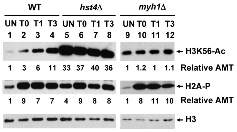
Fig. 3.

The levels of acetylated H3K56 (H3K56-Ac) and phosphorylated H2A (H2A-P) increase following H2O2 treatment in WT cells. S. pombe Hu303, Hu1481 (hst4Δ), and JSP303-Y4 (myh1Δ) were treated with 5 mM of H2O2 for 30 min and then recovered for 0 (T0), 1 (T1), or 3 (T3) hours or remained untreated (UN). Total cell extracts were prepared and separated on two sets of 15% SDS-polyacrylamide gels. Set 1 gel was subject for Western analysis with H3K56Ac antibody. The membrane was then stripped and probed with H2A-P antibody. Set 2 gel was subject for Western analysis with H3 antibody. Lanes 1-8 are in the same membrane while lanes 9-12 are on a separated membrane and Western blotting was performed separately. The amounts of H3K56Ac and H2A-P were quantitated relative to those of H3 (Relative AMT).
