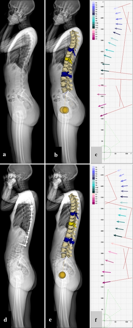
Fig. 3.
Sagittal plane visualizations of the scoliotic spine (a–c) before and (d–f) after correction. Pre-operative and post-operative EOS X-ray images (a, d); images corresponding to pre-operative and post-operative sterEOS 3D reconstructions (b, e); and pre-operative and post-operative full spine vertebra vectors (c, f). Conventional Cobb angulation measurements (shown with red lines) are carried out using vertebra vectors as detailed in “Methods”. See Table 1 for measurement values

