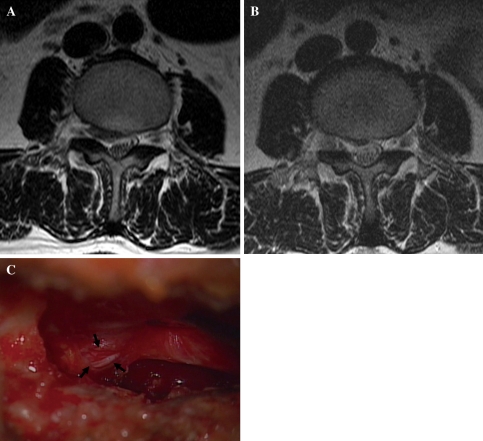
Fig. 3.
Case illustration of unrecognized, nerve root irritation (case No. 5). A 44-year-old female patient underwent transforaminal percutaneous endoscopic lumbar discectomy (PELD) for right-sided disc herniation at the L3–4 level (a). After the procedure, the patient’s pain was relieved, and the postoperative MRI showed a well-decompressed dural sac (b). Seven days later, however, the patient’s leg pain reoccurred and progressed gradually. Open exploration was performed. There was no evidence of recurrent disc herniation. Instead, a small dural defect was found on the operative field. A nerve root was exposed through the dural defect (arrows) and irritated by the surrounding tissues (c)

