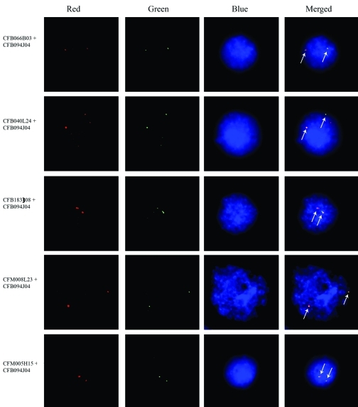
Figure 2.
Double-color FISH showed the six lgbp-containing BAC clones co-localized at the same site on the c. farreri genome. The red, green and blue channels in the pictures were recorded separately and merged to obtain the final figures. The green signals indicate the localization of clone CFB094J04, which was mapped first using single color FISH, whereas the red signals of each set indicate that of the other five clones, respectively. The signals are indicated by arrows in the merged figures. Bar = 5 μm.

