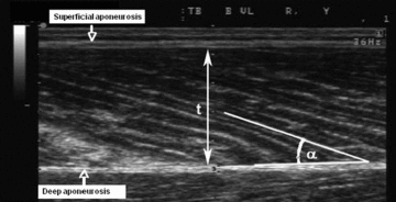
Figure 2. Ultrasound imaging of pennation angle.

Typical ultrasound image of the human vastus lateralis muscle obtained in the sagittal plane using a linear 7.5 MHz probe. Muscle fibres fascicles are clearly visible as the structures stretching from the superficial and deep aponeuroses. t, muscle thickness; α, pennation angle.
