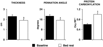
Figure 6. Bed rest effects on vastus lateralis muscle atrophy and oxidative stress.

Values of vastus lateralis thickness, fibre pennation angle and protein carbonylation measured before (Baseline) and after 33 days of bed rest (Bed rest) are shown. Muscle thickness and fibre pennation angle were determined in supine position by ultrasonography approaches. Protein carbonylation was determined by Oxyblot analysis. Oxy RP−1, ratio between quantified oxidized proteins and Red Ponceau stained total protein. §P < 0.001 vs. Baseline; *P < 0.05 vs. Baseline. Statistical analysis was performed by Student's t test.
