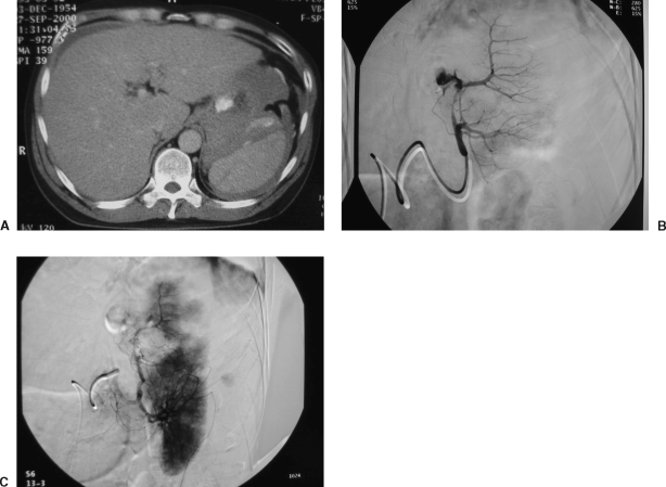
Figure 4.
Splenic embolization in a 45-year-old unrestrained driver in a motor vehicle collision. (A) Contrast-enhanced computed tomography scan demonstrates a large splenic laceration and perisplenic hematoma with active contrast extravasation. (B) Selective splenic angiogram demonstrates active contrast extravasation from the proximal portion of the upper lobe artery. (C) Postembolization angiogram demonstrates cessation of flow to the actively bleeding branch. Note the segmental perfusion abnormalities: these are secondary to Gelfoam embolization. Because of persistent bleeding, a single coil was placed in the proximal bleeding vessel. Note also the uncompromised perfusion to the lower lobe branch distribution.

