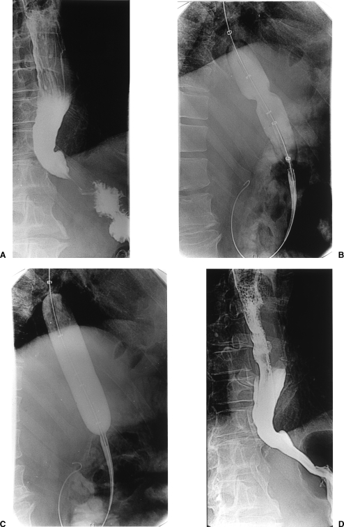
Figure 1.
(A) Contrast esophagogram showing the smooth, tapered beak-like appearance at the level of the esophageal hiatus, characteristic of achalasia. (B) Partially inflated Rigiflex balloon shows a waist at the level of the cardia. (C) Further inflation of the balloon shows complete obliteration of the waist. (D) Barium esophagogram following dilatation shows successful dilation and no evidence of leak.

