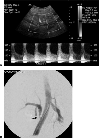
Figure 1.
(A) Ultrasound Doppler examination of the renal artery anastomosis to the external iliac artery demonstrated velocities exceeding 3 m/sec. (B) Selective right external iliac artery angiogram confirms a focal moderately severe and eccentric stenosis at and just distal to the arterial anastomosis (arrow).

