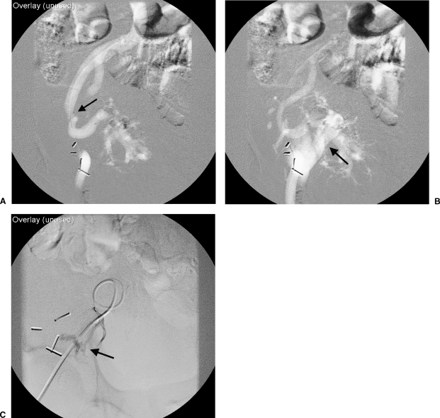
Figure 3.
(A) Carbon dioxide angiogram of the external iliac artery was performed demonstrating focal stenosis (arrow) of the proximal transplant renal artery as demonstrated in Figure 2. (B) Images obtained 0.2 seconds later demonstrate early opacification of the external iliac vein (arrow). (C) Selective injection of a renal segmental arterial branch verifies an arteriovenous fistula (arrow), later corrected with coil embolization.

