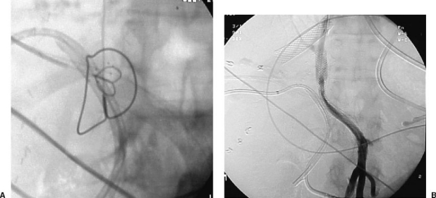
Figure 8.
(A) Lateral spot film demonstrating the gun-sight technique. The small snare is within the portal vein and the large snare within the inferior vena cava. The next step is to perform a puncture with a needle traversing both snares. (B) Portogram after completion of TIPS procedure after using the gun-sight technique. Note that there is a stent within the right hepatic vein. This stent was thrombosed and precluded an optimal hepatic vein access in this case.

