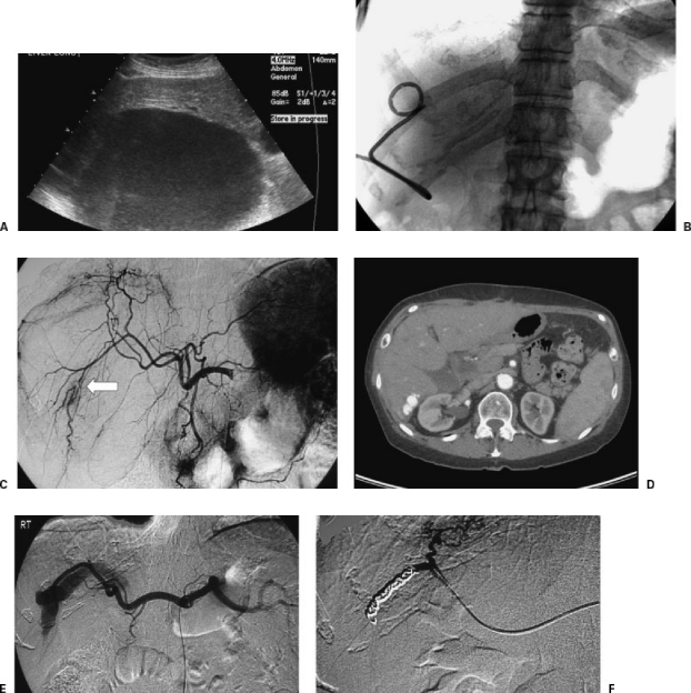
Figure 3.
Arterioportal fistula complicating drainage of a hepatic abscess. A 34-year-old woman with large, hepatic fluid collection presented for drainage. (A) Ultrasonography showed large hepatic fluid collection. (B) Catheter was placed using US guidance without any immediate complication. There was near complete resolution of the cavity by aspiration of the contents. Approximately 15 minutes after catheter placement, the patient became hypotensive and tachycardic. US imaging demonstrated echogenic contents within the previously drained abscess cavity. (C) Angiogram the same day showed segmental interruption of a right hepatic artery branch. In the absence of extravasation, no embolization was performed at this time. (D) Follow-up CT imaging 2 years later showed an arterioportal fistula in the same location. (E) The patient returned to the interventional radiology suite for right hepatic artery angiography, which clearly demonstrated the arterioportal fistula. (F) Following coil embolization, there was cessation of flow to the portal vein.

