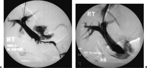
Figure 4.
Portal vein perforation. (A) Direct portogram performed through the Rosch-Uchida TIPS sheath (Cook, Bloomington, IN) demonstrates a large area of extravasation in close proximity to the lower aspect of the main portal vein. A very small inferior mesenteric vein is identified. Note the central puncture site within the main portal vein. (B) Portogram after Wallstent placement still demonstrates the site of extravasation. In this case, placement of a bare stent did not control the portal vein leak and the patient required surgical repair of the portal perforation.

