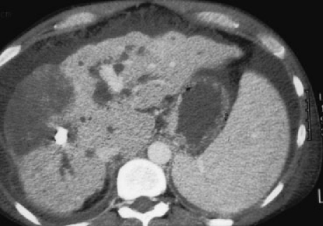
Figure 7.
Liver infarction post–hepatic artery embolization. A contrast-enhanced computed tomography scan demonstrates a low-density, wedge-shaped area in the right lobe of the liver, consistent with a large liver infarction after hepatic artery embolization. Note moderate dilation of the intrahepatic biliary ducts, irregular liver surface, and moderate amount of ascites.

