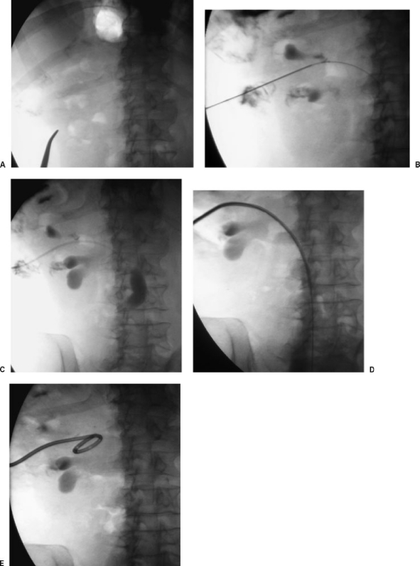
Figure 1.
Percutaneous nephrostomy insertion using single-stick technique. (A) After ultrasound localization, a fluoroscopic image demonstrates overlying ribs and enables appropriate puncture site choice. (B) After ultrasound-guided 21-gauge needle puncture, a small amount of urine is aspirated and contrast is injected. Fluoroscopic image shows a 0.018-inch guide wire is advanced through the needle into the ureter. (C) The needle has been exchanged for coaxial dilators. Fluoroscopic image shows contrast injection into the ureter to confirm position. (D) The 0.018-inch guide wire has been exchanged for a rigid 0.035-inch guide wire and the nephrostomy tube is being inserted over this wire. (E) Final fluoroscopic image shows nephrostomy coiled in renal pelvis.

