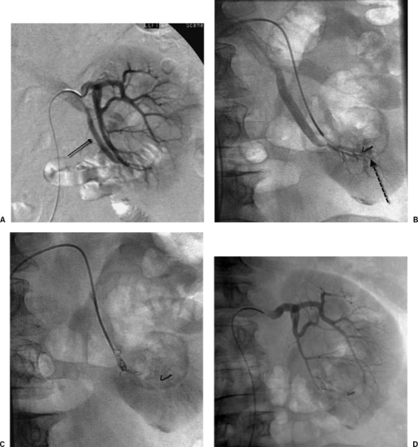
Figure 3.
Embolization of arteriovenous fistula. (A) Selective renal angiogram shows arteriovenous fistula and early draining vein at lower pole of left kidney (small arrow). (B) Subselective digital subtraction angiogram shows arteriovenous fistula at site of wire fragment (dashed arrow). (C) Fluoroscopic image shows coil embolization of lower-pole renal artery. (D) Renal angiogram after embolization shows occlusion of fistula.

