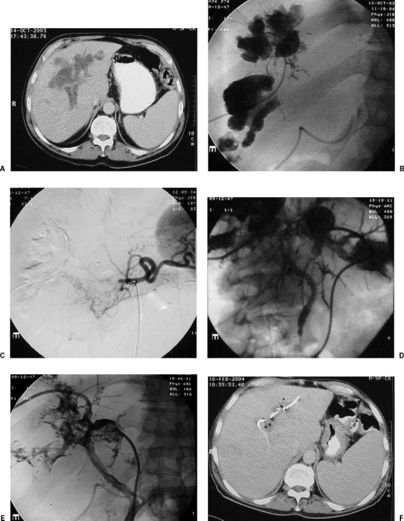
Figure 3.
Patient with hepatic metastasis from a colonic adenocarcinoma consulted us because of ictericia following intra-arterial hepatic treatment performed by means of a reservoir implanted surgically in the proper hepatic artery. (A) The computed tomography (CT) scan showed a liquid collection surrounding the portal branches. The suspected diagnosis was biliary necrosis due to ischemia and chemotoxicity. (B) Image taken after the percutaneous implantation of a catheter in the cavity. The contrast surrounds all the right portal branches and corresponds to a large periportal “biloma.” A catheter can be seen inserted surgically into the hepatic artery. (C) Obstruction of the common hepatic artery with little collateral arterial vascularization to the liver. (D) Two weeks after the placement of two external catheter drainages in both lobes, opacification of small interhepatic biliary branches and of the coledocus can be seen. (E) After the punction of peripheral biliary branches, external-internal catheter drainages are introduced, through the “biloma,” connecting with the coledocus and the duodenum. (F) After great clinical improvement, the CT taken 3 months later shows reduction of the size of the intrahepatic collection and no biliary dilatation.

