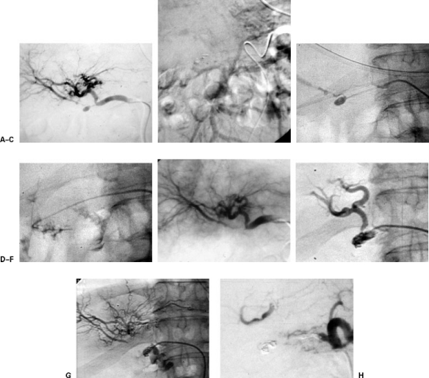
Figure 6.
Patient with pancreatic head carcinoma treated with radiotherapy and surgery. He presents severe digestive hemorrhage. (A) Pseudoaneurysm in right hepatic artery. (B) Indirect portography from splenic artery. Portal thrombosis. (C) Selective catheterization of pseudoaneurysm. (D) Rupture of pseudoaneurysm during the endocavitary manipulation of the coaxial catheter. (E) Arteriography after the selective embolization of the lesion. The flow of the right hepatic artery, through arterial intrahepatic collateras, is preserved. (F) Four months later the patient had another episode of digestive bleeding. In the arteriography a large pseudoaneurysm was observed, placed proximal to the previous one. (G) Further rupture of the pseudoaneurysm during the catheterization. Major digestive bleeding. (H) Arteriography after the complete occlusion of the vascular lesion and the hepatic artery distal and proximal to the rupture. The arterial flow is partially maintained through collaterals of the lesser epiploon. The patient had no further episodes of bleeding or hepatic ischemia.

