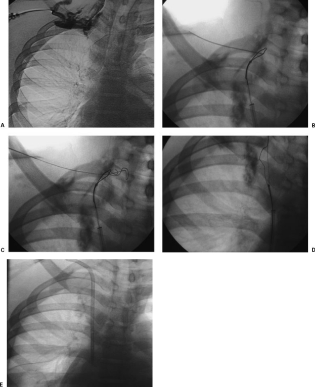
Figure 1.
(A) Venous recannulization in a patient requiring permanent hemodialysis catheter placement. Sonography demonstrated bilateral occlusion of the jugular and subclavian veins. Venography from a right-sided collateral vein verified complete right-sided venous occlusion. (B) A long sheath was placed in the superior vena cava from the femoral venous approach, a glide catheter and wire were used to cross the occluded brachiocephalic vein into a small cervical collateral vein, and a loop snare was opened. This image shows fluoroscopically guided venipuncture using the loop snare as a target. (C) A wire is advanced through the loop snare. (D) The wire is pulled across the occluded veins into the right atrium. (E) A catheter is placed over the wire in standard fashion.

