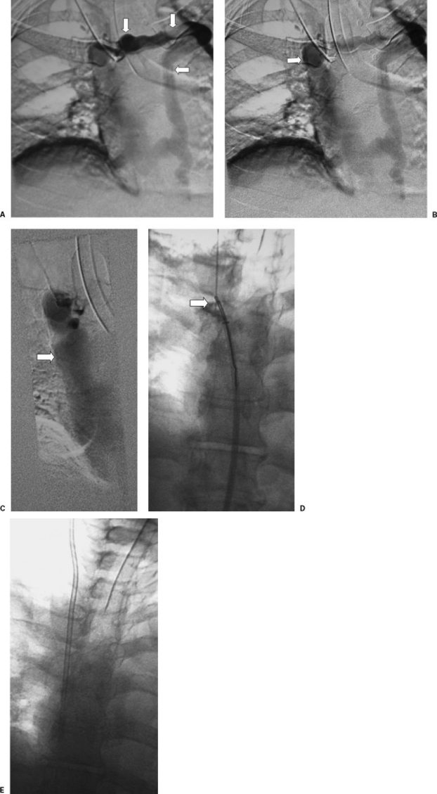
Figure 5.
(A) Sharp venous recannulization in a patient with occluded jugular and subclavian veins. Contrast injection of a cervical vein shows occlusion of the right brachiocephalic vein and filling of a large mediastinal collateral vein (arrows). (B) The collateral vein eventually supplies a dilated azygous arch (arrow). (C) The azygous arch drains into a patent superior vena cava (arrow). (D) Using a 21-gauge needle through a sheath, direct puncture from the cervical collateral vein to a loop snare in the azygous arch was performed. The snare was used to pull a guide wire into the inferior vena cava (arrow). (E) Over the guide wire, a chest catheter was placed in standard fashion.

