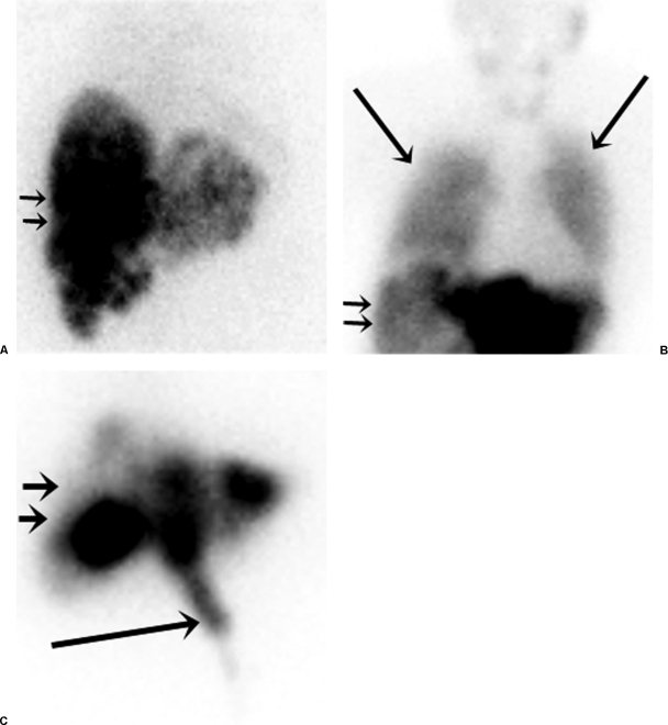
Figure 3.
Nuclear scintigraphy following hepatic arterial injection of Tc99m MAA (liver depicted with small arrows). (A) A 48-year-old woman with intermediate-grade neuroendocrine hepatic metastases demonstrating normal radionuclide distribution with no significant pulmonary or gastrointestinal activity. (B) A 66-year-old woman with hepatic metastatic high-grade neuroendocrine tumor depicting excessive hepatopulmonary shunting (large arrows) that precluded safe treatment. (C) A 55-year-old man with hepatic metastatic colorectal cancer demonstrating gastrointestinal deposition (large arrow) from small unnamed extrahepatic arteries arising from the proper hepatic artery.

