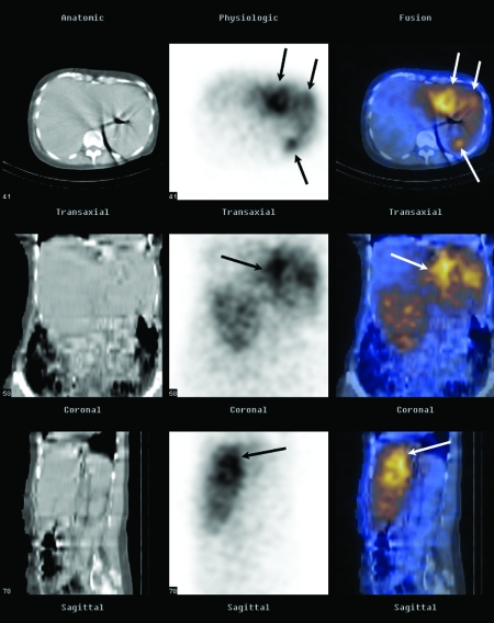
Figure 6.
Bremsstrahlung imaging. Successful targeting of hepatic metastases in a 68-year-old woman (same as Fig. 4). Representative axial, coronal, and sagittal computed tomography (CT) images (first column), single-photon emission computed tomography (SPECT) images (second column), and SPECT/CT fusion images (third column) obtained with a dual-modality imaging system (Hawkeye; GE Medical Systems, Milwaukee, WI) show selective activity in the left hepatic lobe metastases (arrows) ~24 hours after intra-arterial infusion of 35 mCi (945 MBq) of Y90 resin microspheres.

