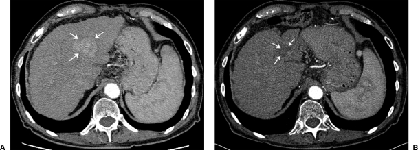
Figure 8.
Hepatocellular cancer. 75-year-old male with hepatitis C–induced cirrhosis and left lobe hepatocellular carcinoma. (A) Pretreatment contrast-enhanced axial CT scan demonstrates a single lesion in the right lobe (arrows). (B) Following a single 1.67 GBq glass microsphere radioembolization (134 Gy dose), a near complete response (arrows) is noted after a 2-year follow-up period.

